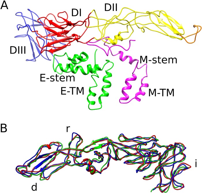
FIG 2.
Structure of the envelope and M proteins on DENV4. (A) Structural organization of the E and M protein heterodimer. Following are the colors of the E protein domains: DI, red; DII, yellow (with the fusion loop in orange); DIII, blue; stem and transmembrane regions, green. The M protein is colored magenta. (B) Superposition of the three individual E proteins in an asymmetric unit. The transmembrane regions are removed for clarity. The most variable parts of the E protein are the loops involved in intradimeric contacts (marked “d”), interraft contacts (marked “r”), and icosahedral 3- and 5-vertex formation (marked “i”).

