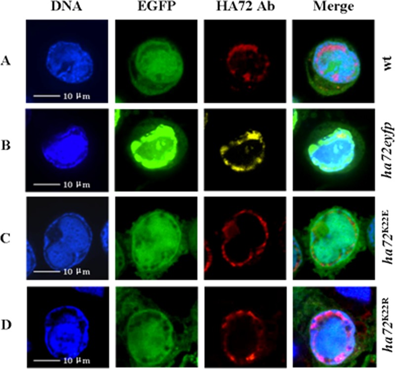
FIG 3.
Subcellular localization of HA72 in HzAM1 cells. Cells were infected with vHaBac-egfp-ph, vHaBacΔ72-72-eyfp-ph, vHaBacΔ72-72K22E-ph, or vHaBacΔ72-72K22R-ph at a multiplicity of infection of 5 and prepared for confocal imaging at 48 hpi. For immunofluoresence assays, polyclonal antiserum against HA72 was used as the primary antibody and stained with anti-rabbit IgG (A, B, and D) or observed via autofluorescence of HA72-EYFP chimera (B). DNA was stained with Hoechst stain. The panels on the right are merged images. Bars, 10 μm.

