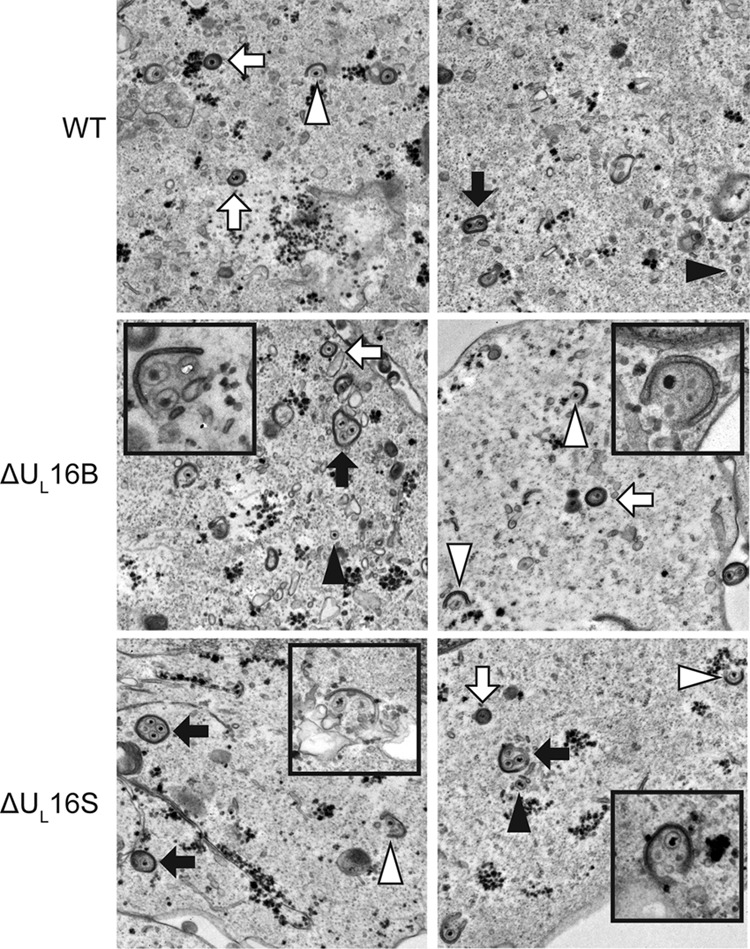
FIG 4.

Multiple capsids are wrapped at once. Representative thin-section electron micrographs of WT- and ΔUL16 mutant-infected (MOI, 1) Vero cells are shown at 24 h postinfection. Simultaneous envelopment of several capsids at a time was detected in ΔUL16 mutant-infected Vero cells (insets). Examples of fully wrapped multicapsid virions (black arrows), single-capsid virions (white arrows), partially wrapped capsids (white arrowheads), and free capsids (black arrowheads) are indicated.
