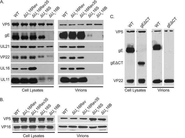
FIG 6.
Cellular expression and packaging of viral proteins. Vero cells were infected with the indicated viruses at an MOI of 5, and the cultures were harvested 18 to 24 h postinfection. Infected cells were directly dissolved in sample buffer (left side of each panel), while extracellular virions were first concentrated by pelleting through a 30% sucrose cushion and then dissolved in sample buffer (right side of each panel). The samples were analyzed by Western blotting with antibodies against the indicated viral proteins, and the amount of each sample loaded was normalized based on the amount of the major capsid protein, VP5. Blots from one of three independent experiments are shown. (A and B) Results for the ΔUL16 mutants and revertant viruses. (C) Results for the mutant lacking the cytoplasmic tail of gE (gEΔCT).

