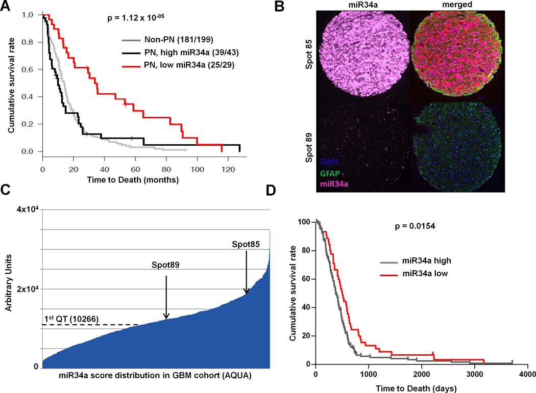
Figure 3. miR-34a levels are prognostic in Glioblastoma.
(A) Kaplan-Meier survival analysis of patients in the TCGA dataset (n=271). Low expression of miR-34a stratifies a subgroup of PN GBM patients with a significant survival advantage compared to others in TCGA cohort (log-rank p=1.12 × 10⁻⁵). (B) GBM Tissue Microarray (TMA). Representative spots showing high and low expressors. A LNA modified probe for miR-34a and and an antibody specific for Glial Fibrillar Acidic Protein (GFAP) were hybridized to the arrays. (C) AQUA score distribution across the GBM cohort. Arrows indicate the AQUA score for the samples shown in figure 3B. (D) Kaplan-Meier survival analysis of patients in the independent training cohort (n=220). Subjects were stratified in high and low miR-34a expressors based on the quantile expression assessed by the AQUA score. 1st (lower) quartile of the AQUA score distribution was used as cut-off to assign patients to the low or high expressors groups. “miR-34a low” patients show a significant better outcome when compared to “high expressors” (p=0.0154).

