Figure 3.

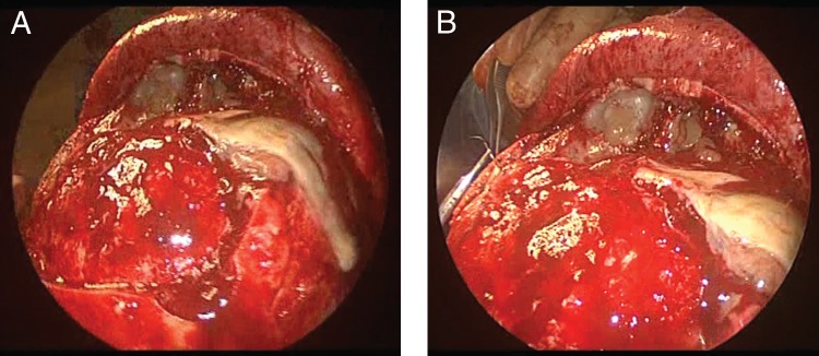
Patient 1. Intraoperative photographs of (A) bifrontal craniotomy with (B) dural repair. Note mucopurulent drainage observed on exposure of the frontal sinus.

Patient 1. Intraoperative photographs of (A) bifrontal craniotomy with (B) dural repair. Note mucopurulent drainage observed on exposure of the frontal sinus.