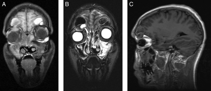
Figure 5.
Preoperative (A) coronal T1-MRI postcontrast, (B) coronal T2-MRI, and (C) sagittal T1-MRI postcontrast show bilateral expansile lesions of variable density within the frontal sinuses measuring 2.3 cm on the right and 2.1 cm on the left. Extension into the bilateral anterior cranial fossa and the superior aspect of the right orbit are present with inferior displacement of the right globe. MRI, magnetic resonance imaging.

