Figure 3.
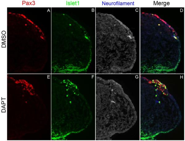
Notch inhibition leads to premature neuronal differentiation in the ectoderm. Transverse sections through the ophthalmic trigeminal (opV) placode region of a 20-22 ss embryo. The heads were cultured in either DMSO or DAPT for 12 hrs, harvested, cryosectioned, and immunostained for the opV marker Pax3 (A,E), the early neuronal marker Islet1 (D,F), and the late neuronal marker neurofilament (C,G). DMSO treated embryos showed normal opV placode development (A-D). DAPT treated embryos showed an increase in ectodermal Pax3+cells coexpressing Islet1, and the number of Pax3+/Islet1+ mesenchymal cells was also dramatically increased (E,F,H). DMSO and DAPT treated embryos showed no neurofilament expression in the ectoderm or in the mesenchyme (C,G).

