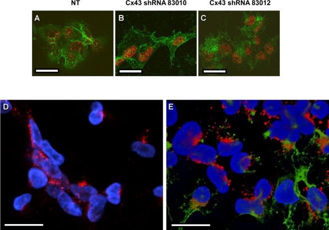
FIG 3.
Analysis of VacA and Cx43 localization. (A to C) AZ-521 cells expressing nontargeting shRNA (NT), Cx43-specific shRNA 83010, or Cx43 shRNA 83012 were incubated with 5 μg/ml of acid-activated Alexa 488-labeled VacA in the presence of 10 mM ammonium chloride at 37°C for 1 h. Cells were imaged as described in Materials and Methods. The nucleus is shown in red, and VacA localization is shown in green. (D and E) AZ-521 cells were incubated with 5 μg/ml of acid-activated VacA in the presence of 10 mM ammonium chloride at 37°C for 1 h, and control cells were not treated with VacA. Cells were fixed, permeabilized, and stained to detect VacA and Cx43, as described in Materials and Methods. Panel D shows AZ-521 control cells that were not treated with VacA, and panel E shows AZ-521 cells treated with VacA for 1 h. The nucleus is shown in blue, Cx43 is shown in red, and VacA is shown in green. Bars, 20 μm.

