FIG 4.
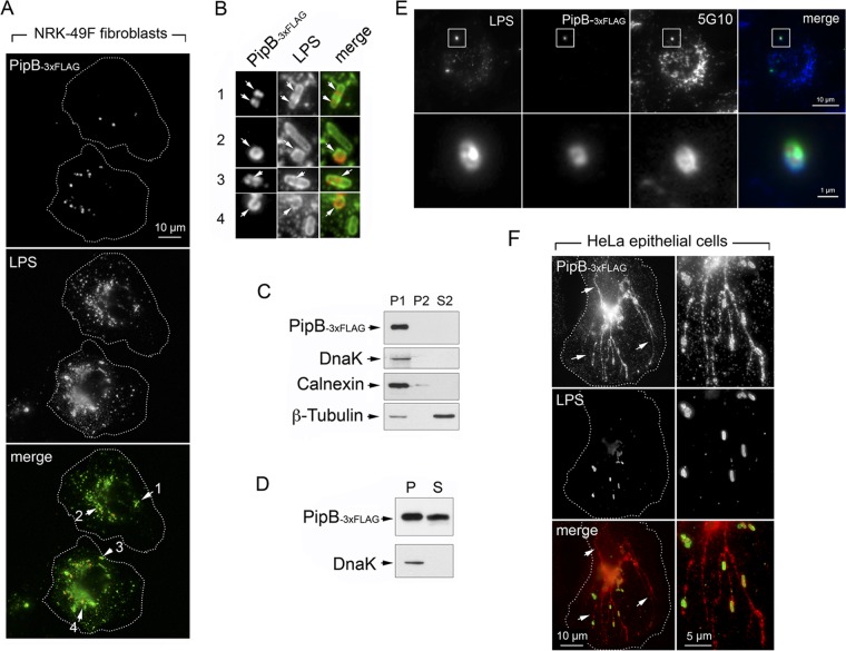
SPI-2 effector PipB is translocated by nongrowing dormant intracellular S. Typhimurium to phagosome-confined regions. (A) Distribution of endogenous chromosomally tagged PipB-3×FLAG in NRK-49F fibroblasts at 24 h postinfection. Arrows indicate four fields containing intracellular bacteria that are magnified in panel B. (B) Magnified images of fields selected from panel A showing the distribution of PipB-3×FLAG in dormant intracellular bacteria. Note the lack of effector labeling in some bacteria. (C) Mechanical fractionation denotes that endogenous PipB-3×FLAG is not released to the endomembrane system. P1, nuclear/bacterial fraction; P2, high-speed particulate material, membranous fraction; S2, cytosol, soluble fraction (see Materials and Methods for details). (D) Translocation of PipB-3×FLAG by nongrowing dormant intracellular bacteria shown by the detection of the effector in a subcellular fraction containing material solubilized with 1% (vol/vol) Triton X-100. P, pellet, material insoluble in detergent; S, material solubilized with the detergent. (E) Triple labeling showing that the distribution of PipB-3×FLAG in NRK-49F fibroblasts is observed inside that of a membrane marker positioned in the phagosomal membrane, a glycoprotein recognized by the mouse monoclonal 5G10 (64). (F) Distribution of endogenous PipB-3×FLAG in HeLa epithelial cells at 6 h postinfection. Note that PipB shows a distribution confined to the phagosomal region exclusively in nongrowing dormant bacteria located inside fibroblasts.

