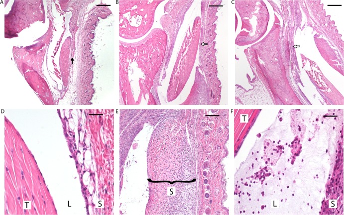
FIG 3.
(Top row) Differences in tibiotarsal changes in C3H mice infected with B. burgdorferi clones at 2 weeks postinfection. (A) ΔArp. (B) Wild type. (C) cArp. Reduced cellular infiltration and synovial hyperplasia (black arrow versus white arrows) and minimal to absent neutrophilic infiltration were observed in the tibiotarsal flexor tendons of mice infected with the ΔArp clone. Histopathological changes between the WT and cArp clones were indistinguishable. (Bottom) High-power magnification of histopathologic changes in C3H mice infected with B. burgdorferi clones. (D) Minimal changes observed in the tibiotarsal flexor tendon (T) of ΔArp clone-infected mice with markedly reduced synovial hyperplasia and little to no neutrophilic infiltration in the lumen (L) of the tendon sheath (S) (magnification, ×100). (E) Inflamed hyperplastic tendon sheath in a mouse infected with wild-type B. burgdorferi (magnification, ×60). (F) Synovial lumen with inflammatory exudate comprised of neutrophils, edema residue, and fibrin and hyperplasia of the tendon sheath observed in mice infected with the cArp clone (magnification, ×100).

