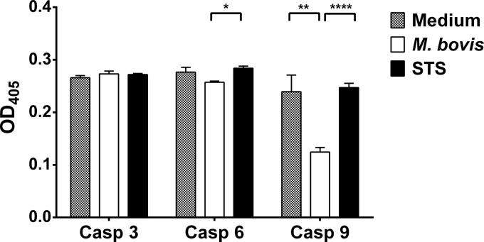
FIG 3.
Caspase activity of M. bovis-infected monocytes. Data for the activation of caspases 3, 6, and 9 in cytosolic fractions of monocytes after 24 h of incubation with M. bovis are shown. Activation is based on the cleavage of the corresponding synthetic substrates (caspase 3, DEVD; caspase 6, VEID; caspase 9, LEHD) labeled with para-nitroaniline, which is released after the substrate is cleaved by the specific caspase. All data were square root transformed before analysis and were generated from 3 animals and triplicate cultures for each treatment. Each bar represents the mean of 3 replicates for each treatment, and the error bars indicate the standard deviations from the means of each treatment. The x axis shows specific caspases (caspases 3, 6, and 9), and the y axis indicates the optical density at 405 nm (OD405) of the samples. Significant differences for each caspase activity between the treatments are indicated by ∗ (P ≤ 0.05), ∗∗ (P ≤ 0.01), and ∗∗∗∗ (P ≤ 0.0001).

