
FIG 3.
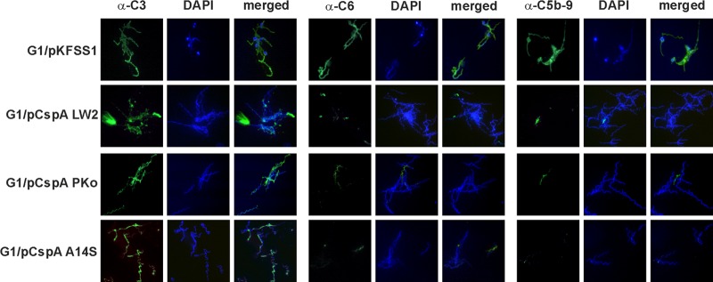
Deposition of complement components C3, C6, and TCC on the surface of spirochetes. Complement components deposited on the surface of transformants G1/pKFSS1, G1/pCspA LW2, G1/pCspA PKo, and G1/pCspA A14S were visualized by indirect immunofluorescence microscopy. Spirochetes (6 × 10⁶) were incubated with 25% NHS for 30 min at 37°C with gentle agitation, and bound C3, C6, and C5b-9 (TCC) were analyzed with specific antibodies against each component and appropriate Alexa Fluor 488-conjugated secondary antibodies. For visualization of the spirochetes in a given microscopic field, the DNA-binding dye 4′,6-diamidino-2-phenylindole (DAPI) was used. The spirochetes were observed at a magnification of ×100. The data were recorded with an Axio Imager M2 fluorescence microscope (Zeiss) equipped with a Spot RT3 camera (Visitron Systems). Each panel is representative of at least 20 microscope fields.