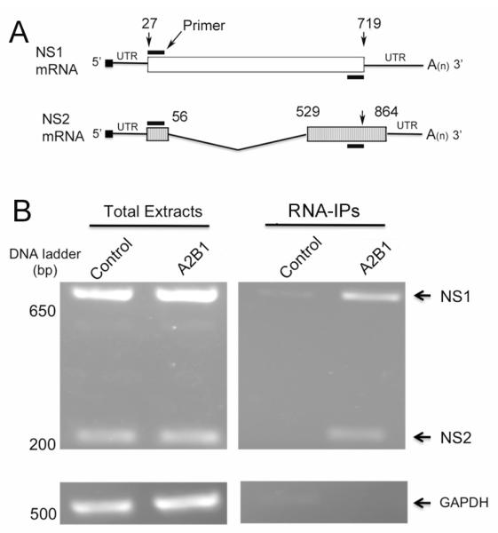
Fig. 5.
hnRNP A2/B1 proteins are associated with NS1 and NS2 mRNAs. (A) Schematic representation of NS1 mRNA, NS2 pre-mRNA and the positions of the primers used to amplify NS1 and NS2 mRNAs. The coding regions of NS1 and NS2 mRNAs are shown as white and hatched boxes, respectively. The numbers above the coding regions indicate the start and end nucleotide positions in the NS1 and NS2 mRNAs. The NS2 mRNA is alternatively spliced from NS1 mRNA, and the V-shaped line denotes the region that is removed in splicing. With the primers (black bar) indicated in the diagram, the amplicon size for NS1 and NS2 was calculated to be 693 and 221 bps, respectively. (B) hnRNP A2/B1 proteins are associated with NS1 and NS2 mRNAs. 293T cells transiently transfected with the plasmids that express HA-hnRNP A2/B1 or HA tag alone (control) were infected with A/PR/8/34 viruses at an MOI of 3. At 10 hpi, the cells were harvested and lysed, and the resulting whole cell lysates were immunoprecipitated with immobilized anti-HA antibody. The RNAs immunoprecipitated were released from the complexes by incubating with proteinase K, reverse-transcribed, and PCR-amplified with the primers indicated in (A). The resulting DNAs were examined by a 1.3% agarose gel. GAPDH was used as an internal reference.

