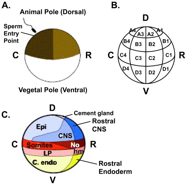
Figure 3.1. Fate map of a 32-cell Xenopus laevis embryo.
(A) Following fertilization and cortical rotation, cells derived from the lightly pigmented side of the embryo will form rostral (R) structures and the darkly pigmented side will give rise to more caudal (C) structures. This schematic represents a 90° rotation from the traditional dorsal/ventral (D/V) axis. It should be noted that neither the original fate map, nor the updated version is strictly accurate in assigning the D/V axis since dorsal and rostral fates are closely aligned, as are ventral and caudal fates. (B) Blastomere nomenclature of a 32-cell (stage 6) embryo (only cells in one of two sides with respect to the left-right axis are shown). (C) Prospective adult tissues derived from regions of a 32-cell embryo, as seen from a side view. C). Adapted with permission from (21). D = Dorsal, V = Ventral, R = Rostral, C= Caudal, Epi = Epidermis, CNS = Central Nervous System, No = Notochord, LP = Lateral Plate Mesoderm, hm = head mesoderm, endo = endoderm.

