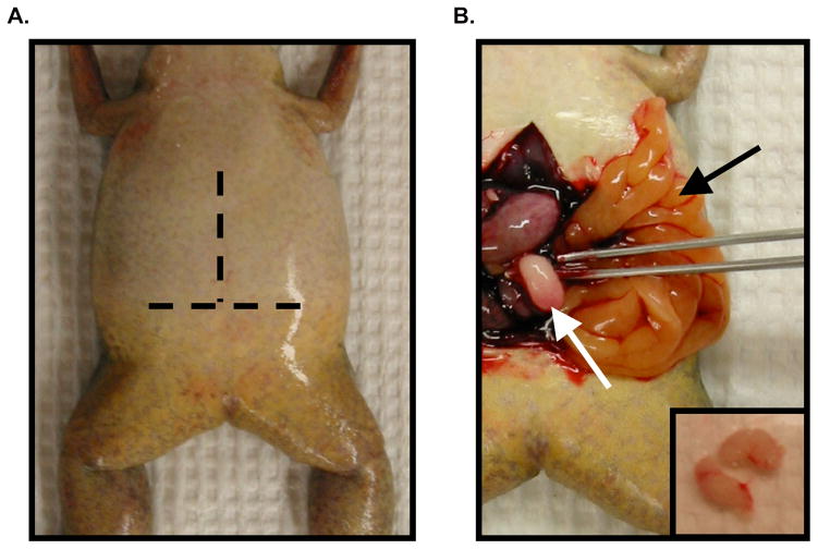
Figure 3.3. Harvesting testes from a male frog.
(A) Ventral view of an anesthetized male frog. Dashed lines represent the approximate location of horizontal and vertical incisions to be made. (B) Testis removal following incisions in panel A and exanguination (described in Methods). The testis (white arrow, and inset) is located at the end of the fat bodies (black arrow) on the left and right side of the abdominal cavity.

