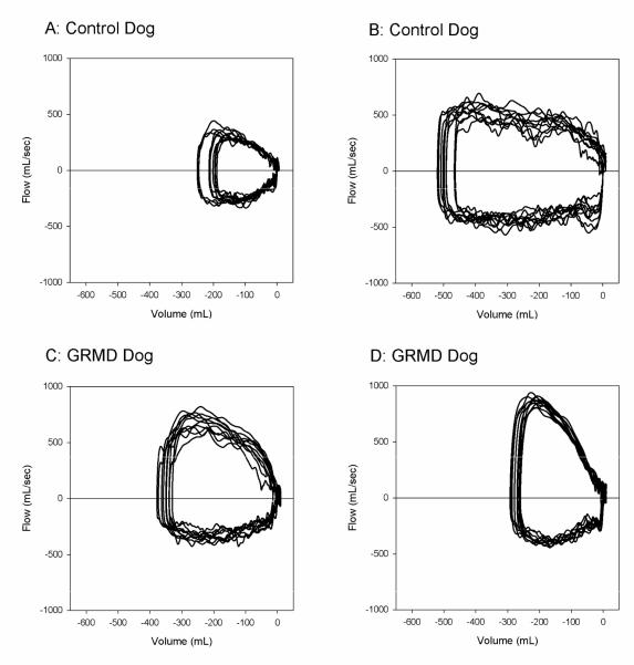
Figure 1.
Tidal breathing flow-volumes (F-V) loops of 10 representative breaths from two control dogs (A, B) and two GRMD dogs (C,D) displayed on graphs with identical scales. These dogs were selected as representative of their groups based on peak tidal expiratory flows that were closest to the median value for their group. The loops from GRMD dogs show visibly greater expiratory flows (positive values) compared with inspiratory flows (negative values) and compared with expiratory flows from control dogs.

