
Figure 7.
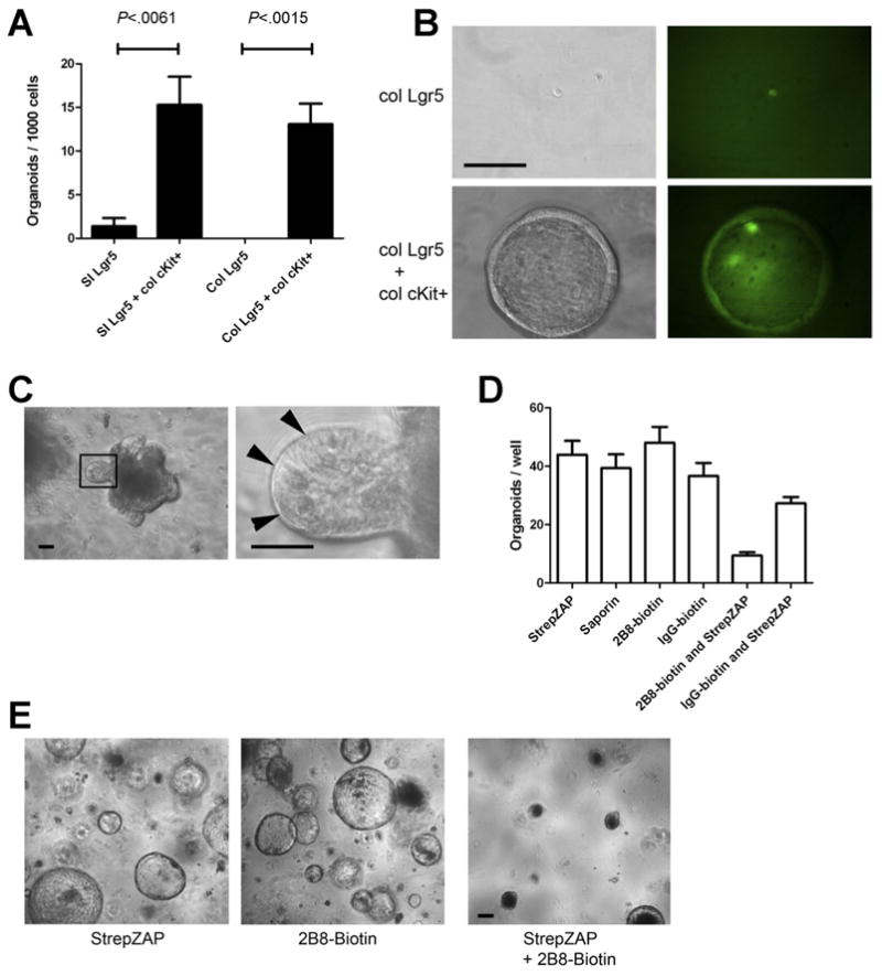
cKit+ cells promote organoid formation of Lgr5+ cells. (A) Lgr5-GFP+ cells were FACS isolated from the proximal small intestine or colon and were cultured alone, with FACS-sorted colonic cKit+CD44+ epithelial cells, or with cKit−CD44− epithelial cells. Organoids were scored 7 days after plating. Histogram shows organoids per 1000 FACS-sorted cells. n = 3. P values indicated. Error bars indicate standard error of mean. (B) Representative images at 1 week post plating from Lgr5-GFP colonic cells grown alone (top rows) or with CD44+cKit+ colonic cells (bottom rows). Left column shows phase contrast images, and right shows GFP. (C) Primary small intestinal organoids (left) contained visible Paneth cells (right). (D) Organoids shown in (C) were passaged by dissociation into a single-cell suspension and were treated as shown. StrepZAP is streptavidin-saporin conjugate. n = 3. (E) Representative images from (D) are shown. Images acquired 7 days after passaging. Scale bars: 50 uM.