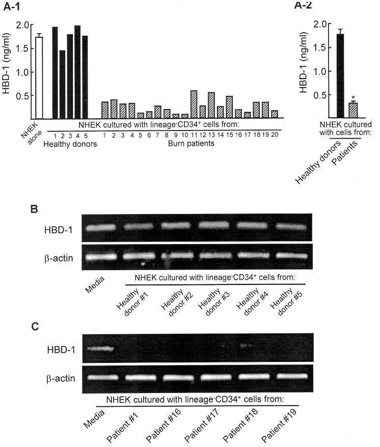
Figure 1. HBD-1 production and mRNA expression by NHEK cultured with peripheral blood lineage−CD34+ cells that were isolated from both severely burned patients and healthy donors. A.
HBD-1 production. Lineage−CD34+ cells (1×10⁵ cells/ml, upper chamber), isolated from 5 healthy donors (#1∼#5) and 20 burn patients (#1∼#20), were transwell-cultured with NHEK (1×10⁵ cells/ml, lower chamber) for 36 hours. After removal of the upper chamber, NHEK in the lower chamber were cultured for an additional 36 hours. Culture fluids obtained were assayed for HBD-1 by ELISA. Fig. 1A-1 shows independent experiments performed using blood specimens from 5 healthy donors and 20 burn patients, and Fig. 1A-2 shows mean ± SEM of the results shown in Fig. 1A-1. * P<0.001 vs control. B and C. HBD-1 mRNA expression. Lineage−CD34+ cells (1×10⁵ cells/ml, upper chamber), isolated from peripheral blood of 5 healthy donors (#1∼#5, B) and 5 severely burn patients (#1, #16, #17, #18, #19, C), were transwell-cultured with NHEK (1×10⁵ cells/ml, lower chamber) for 36 hours. After removal of the upper chamber, NHEK in the lower chamber were analyzed for HBD-1 mRNA by RT-PCR.

