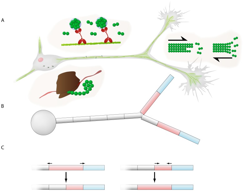
Figure 1. Illustration of the neurite outgrowth model.
(A) Tubulin dynamics in the model. Tubulin molecules (green spheres) are produced in the soma, in biological neurons via translation of mRNA on ribosomes (the brown structure). Tubulin is then transported by diffusion and active transport; in biological neurons, the microtubule bundles (the light green fibers) act as railway tracks on which the tubulin molecules are bound via motor proteins (the red molecules). Tubulin is transported to the growth cones at the tip of the neurites. At the growth cone, tubulin is integrated or polymerized into the microtubule cytoskeleton (long polymers of tubulin dimers; the green fibers), which elongates the neurite. When the microtubule depolymerizes, the neurite retracts and tubulin becomes free again. (B) The neuron is divided into multiple compartments. The soma is represented by a single compartment; it connects to a number of neurites consisting of a series of connected compartments. The compartment at the tip of the neurite represents the growth cone (blue). To minimize artificial fluctuations in tubulin concentration, the growth cone is moved forward during growth but its size remains constant, while instead the second compartment (pink) is elongated. (C) The elongating compartment dynamically splits if it becomes too large; likewise, if a shrinking compartment becomes too small, it merges with its proximal (parent) compartment.

