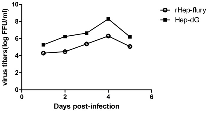
Figure 3. Growth kinetics of HEP-dG and rHEP-Flury in BHK-21 cells.

Cells were infected with HEP-dG (▪) and rHEP-Flury (○) at a MOI of 5. The cultures were incubated at 37°C. Supernatants were harvested at 1, 2, 3, 4 and 5 days post inoculation, and virus titers examined by the fluorescent-focus assay.
