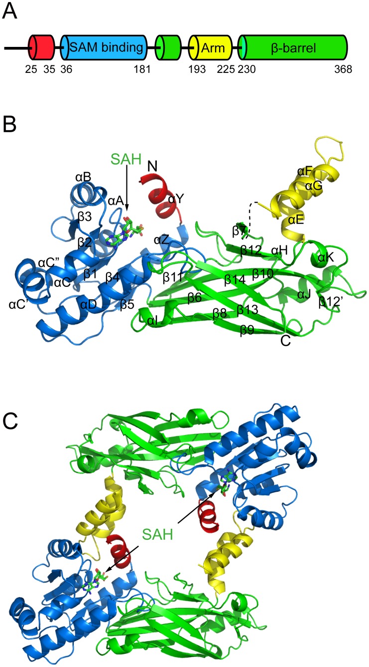
Figure 1. Structure of TbPRMT6.
(A) Schematic diagram of the domain arrangement of TbPRMT6 (B) Overall structure of a monomer The N-terminal helix αY, the SAM-binding domain, the dimerization arm, and the β-barrel domain are shown in red, blue, yellow, and green, respectively The cofactor SAH is shown in the stick model The segment between helix αG and strand β7 is invisible and is shown as a dashed line (C) Structure of the TbPRMT6 dimer.

