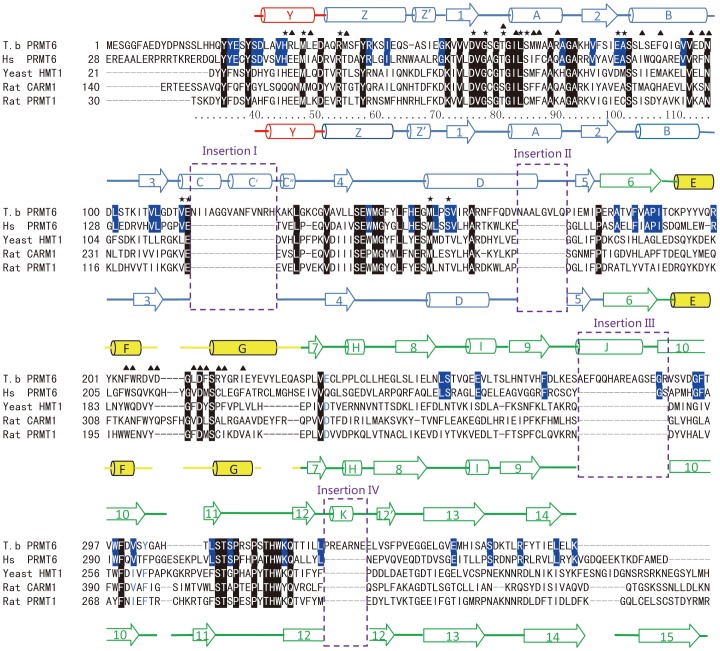
Figure 3. Sequence alignment of TbPRMT6, human PRMT6, rat PRMT1, and rat CARM1.
The secondary structural elements of TbPRMT6 and rat PRMT1 are shown on the top of and underneath the sequence, respectively The color of the secondary structural elements is the same as in Figure 1B The residues conserved among the four enzymes are highlighted in black, and the residues conserved in the PRMT6 paralogs are highlighted in blue The asterisks and triangles above the sequence indicate the residues in TbPRMT6 that are involved in SAH recognition and dimerization, respectively The four stretches of insertions are bracketed in purple dashed frames and labeled.

