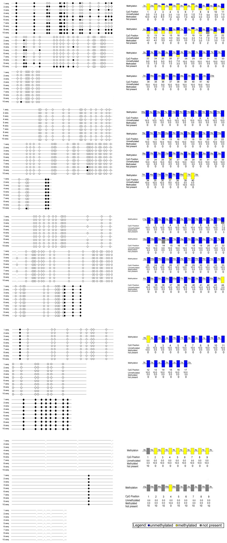
Figure 3. Methylation status of the β-catenin promoter region in SPC.
Mapping of the BSP results of lung cancer cell lines SPC, showing the methylation status of the β-catenin promoter region. The filled circles represent methylated CG sites, hollow circles represent unmethylated CG sites. Yellow indicates methylation, blue indicates unmethylated, gray indicates no CG site.

