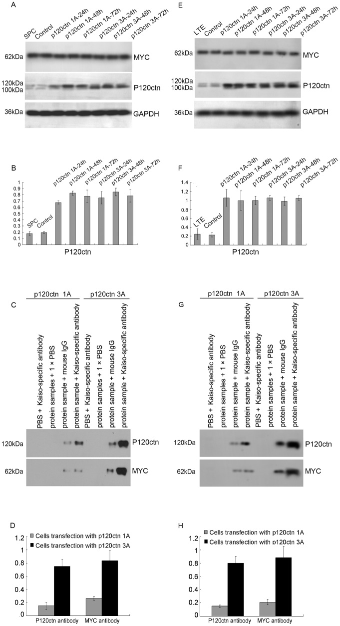
Figure 6. The binding of p120ctn isoforms 1A and 3A with Kaiso.
We introduced plasmids encoding DDK-MYC tagged p120ctn isoforms 1A and 3A cDNA into the lung cancer cell lines, and verified the effect of transfection by Western blot using p120ctn and MYC specific antibodies. P120ctn specific bands were detected at 120 kDa and 100 kDa and MYC specific bands were detected at 62 kDa in following transfection of the SPC (A) and LTE (E) cell lines. Statistical analysis by t-test in SPC (B) showed that cells transfection with p120ctn-1A (P<0.001 for 24 h, P<0.001 for 48 h, and P = 0.007 for 72 h, respectively) and -3A (P = 0.008 for 24 h, P = 0.001 for 48 h, and P = 0.008 for 72 h, respectively) showed significant expression, compared with control cells. Similar results to those in the SPC cell line were seen in the LTE line (F: in the cells transfection with p120ctn-1A, P = 0.013 for 24 h, P = 0.023 for 48 h, and P = 0.001 for 72 h, respectively; in the cells transfection with p120ctn-3A, P<0.001 for 24 h, P = 0.002 for 48 h, and P<0.001 for 72 h, respectively). Co-immunoprecipitation results confirmed that Kaiso formed complexes with proteins expressed by p120ctn isoform plasmid transfected SPC (C) and LTE (G) cell lines, and the statistical analysis by t-test in SPC (D) and LTE (H) showed that the binding ability of kaiso with p120ctn isoform 1A was significantly less than that of p120ctn isoform 3A (D: P<0.001 for p120ctn, P = 0.002 for MYC, respectively; H: P<0.001 for p120ctn, P<0.001 for MYC, respectively).

