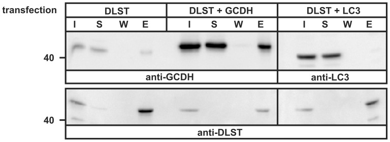
Figure 3. Co-Precipitation of DLST with GCDH.
Extracts from HeLa cells overexpressing the DLST-His6 alone (DLST) or together with GCDH-Myc (DLST+GCDH) were incubated with Ni-NTA agarose for 4 h. Aliquots of the cell extract (input, I: 10% of total), the unbound protein supernatant after precipitation of Ni-NTA beads (S, 10%), the last wash (W, 25%) and the eluted fraction (E, 100%) representing bound proteins, were analyzed by successively exposing the blot to anti-GCDH and, after stripping, to anti-DLST antibodies. Extracts of HeLa cells overexpressing DLST-His6 and LC3-GFP (DLST+LC3) were used as negative control and analyzed by anti-LC3 western blotting. The expression of DLST was analyzed by anti-DLST western blotting. The position of the 40 kDa molecular mass marker protein is indicated. The figure shows a representative blot of n = 3 independent experiments.

