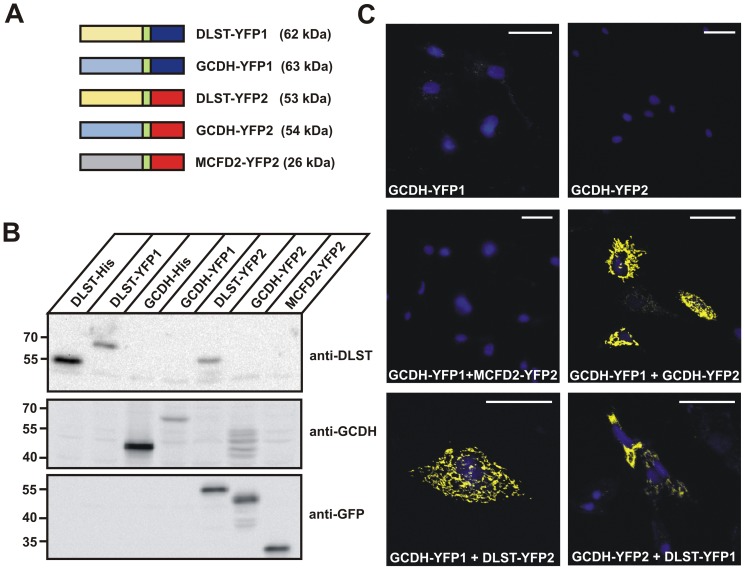
Figure 5. YFP fragment complementation assay demonstrates the interaction of GCDH with DLST in vivo.
(A) Schematic composition of C-terminal YFP1 (dark blue) and YFP2 (red) fusion proteins of DLST, GCDH and MCFD2 used in this study. The 10-amino acid linker (GGGGS)2 is indicated in green. The calculated molecular masses of the fusion proteins are shown in brackets. The ERGIC marker protein MCFD2-YFP2 was used as negative control. (B) Expression analysis in BHK cells of all fusion proteins visualized by western blotting, using anti-DLST, anti-GCDH and anti-GFP antibodies. (C) Fluorescence microscopy of the indicated single or co-expressed fusion proteins. Strong YFP fluorescence was observed in cells co-expressing either GCDH-YFP1 with DLST-YFP2 or GCDH-YFP2 with DLST-YFP1. Nuclei were visualized using DAPI (blue). Scale bars = 40 µM. Representative images of n = 3 independent transfection experiments are shown.

