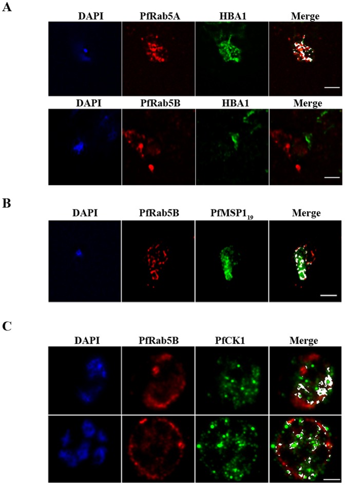
Figure 5. PfRab5B colocalises with PfMSP1 and PfCK1, but not with haemoglobin.
(A) PfRab5A colocalises with haemoglobin (HBA1) containing vesicles (r = 0.724), unlike PfRab5B (r = 0.081; n = 3). (B) PfRab5B colocalises to differing degrees (r = 0.849; n = 3) with the C-terminal 19 kDa fragment of PfMSP1 on structures close to the food vacuole and the parasite nucleus shown in blue by DAPI staining. (C) PfRab5B colocalises with PfCK1 on intracellular structures (r = 0.615; n = 3) and at the parasite plasma membrane (r = 0.475; n = 3). Areas of colocalistaion are shown in white and used to calculate Pearson's r coefficients. Scale bars, 2 µm.

