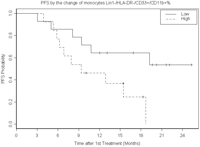
Figure 3. Kaplan-Meier plot of progression free survival (PFS) by the dichotomized change in the percentage of circulating MDSC between baseline and week 6.
Greater decrease in circulating monocyte gate MDSC (Lin1−/HLA-DR−/CD33+/CD11b+%) was associated with improved progression free survival (PFS; p = 0.03; N = 27 patients). Example of raw data is provided in Figure S4 where the gating strategies used for MDSC subsets are shown.

