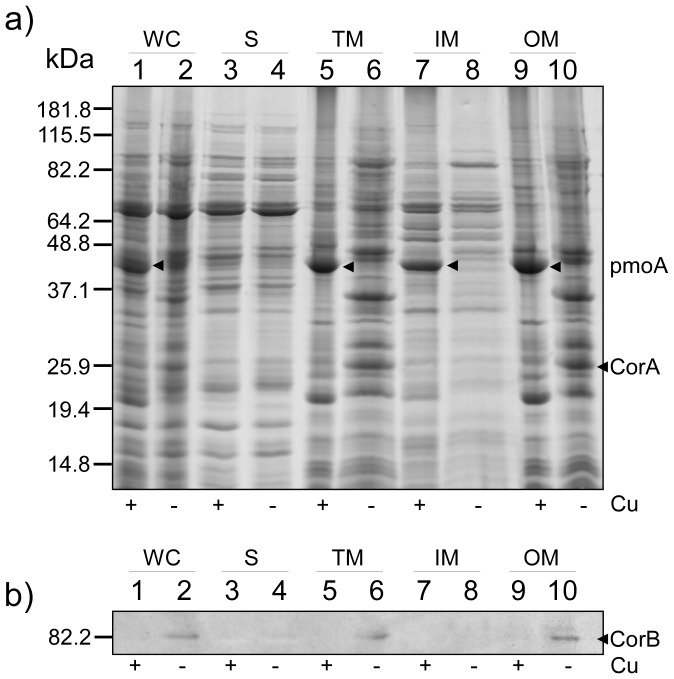
Figure 1. SDS-PAGE and protein immunoblot analysis of proteins obtained during the fractionation of high- and low-copper grown M. album BG8.
Samples from each fractionation step were collected and comparable amounts were analyzed. a) A 12.5% PA-gel was used and stained with Coomassie Brilliant Blue R-250. a) and b) Lane 1 and 2, whole cells (WC); lane 3 and 4, soluble fraction (S); lane 5 and 6, total membrane fraction (TM); lane 7 and 8, Triton X-100 soluble membrane fraction (enriched inner-membrane fraction, IM); lane 9 and 10, Triton X-100 insoluble fraction (enriched outer membrane fraction, OM). High- (+) and low-copper (−) conditions during growth are indicated below the PA-gel. b) Protein immunoblot of a) using CorB-specific antibody [28]. CorA and putatively the pmoA subunit of the M. album BG8 pMMO are indicated with arrowheads. Molecular mass markers are indicated to the left of both a) and b).

