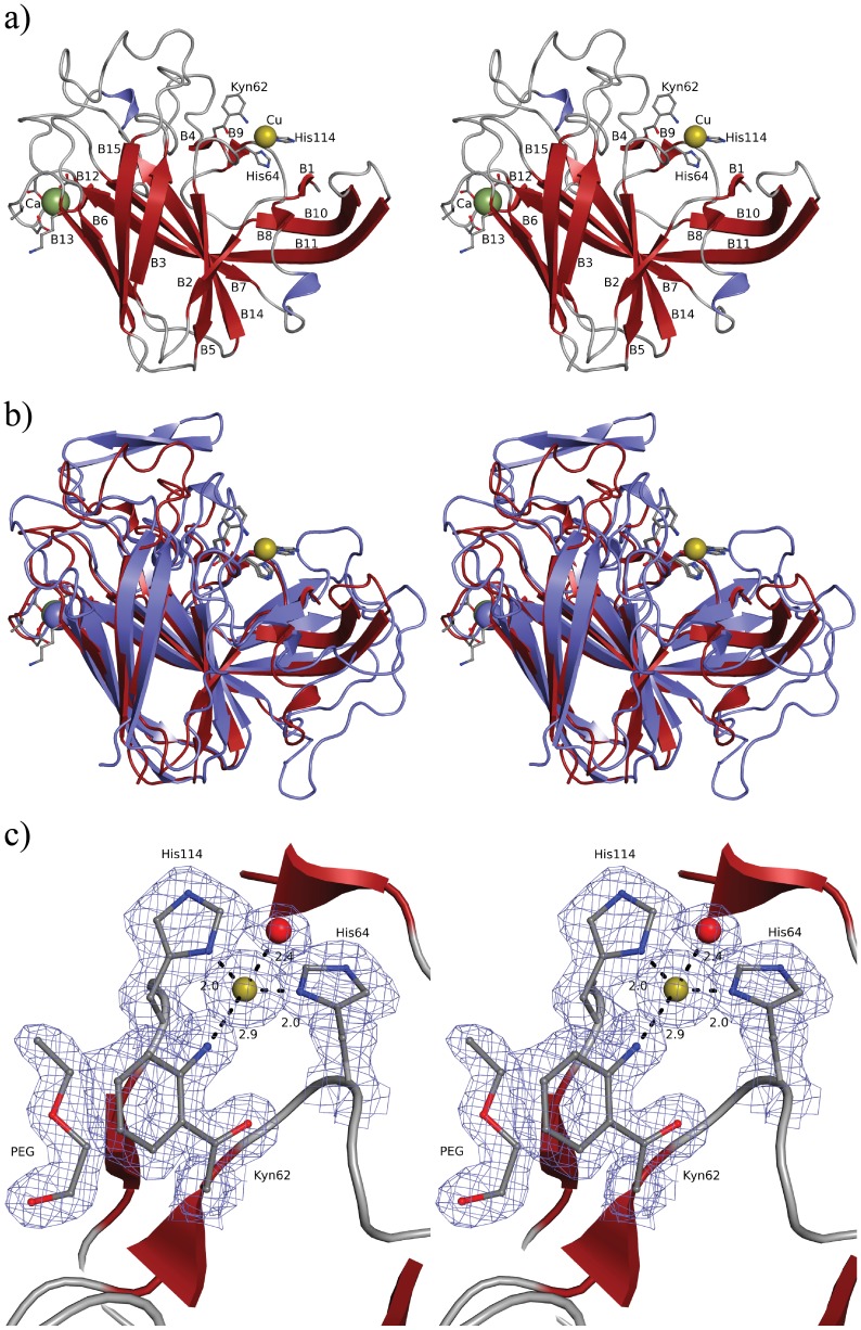
Figure 6. Overall view and copper-binding site of CorA.
a) Stereo plot of CorA, illustrating the 15 β-strands, Cu(I)(yellow sphere), Ca2+ (green sphere), and residues coordinating the metal ions (ball-and-stick models). b) Superimposition of CorA (red) and MopE* (blue). The copper and calcium in CorA are illustrated as yellow and green spheres, respectively. The metals in MopE* are coloured blue. Because of the superposition calcium appears as blue and copper as yellow. The 15 β-strands of CorA superimpose well on 15 of the 21 strands found in MopE*. Also the residues involved in copper and calcium binding superimpose well. c) Stereo plot illustrating the 2foDfc electron density, contoured at 1 σ in the copper binding site, demonstrating the oxidation of tryptophan to kynurenine at residue 62. The copper and the coordinating water are illustrated as yellow and red spheres, respectively. The polyethylene glycol molecule shown is found near the kynurenine residue in all CorA molecules in the crystal structure.

