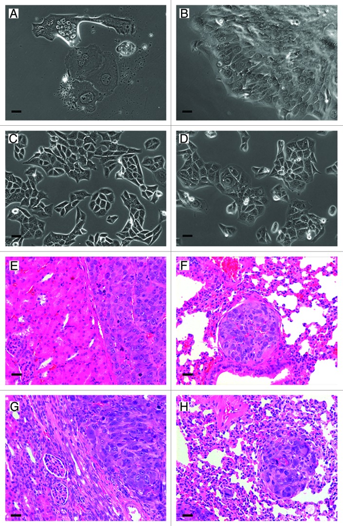
Figure 4. Docetaxel-resistant cells cultured in vitro and after transplantation. Morphological appearance of docetaxel-resistant cells one week post-isolation from docetaxel treated macrobeads (A). Maintained in vitro for 16–17 weeks docetaxel-resistant cells developed plaques (B). These cells were passaged at week 16 post-treatment, and within 2 weeks the harvested cells assumed normal RENCA morphology (C). As a reference, RENCA monolayers demonstrating normal RENCA cell morphology (D). Representative H&E stained tissue of a Balb/cJ mouse at 56 d post-transplantation shows tumor formation inthe left kidney (E) and lung metastasis (F). Representative H&E stained tissue from tumor formation in the left kidney (G) and lung metastasis (H) of a NOD.CB17-Prkdcscid/J mouse at 66 d post-transplantation. Original magnification for all panels = 200×, scale bar: 40 µm.

An official website of the United States government
Here's how you know
Official websites use .gov
A
.gov website belongs to an official
government organization in the United States.
Secure .gov websites use HTTPS
A lock (
) or https:// means you've safely
connected to the .gov website. Share sensitive
information only on official, secure websites.
