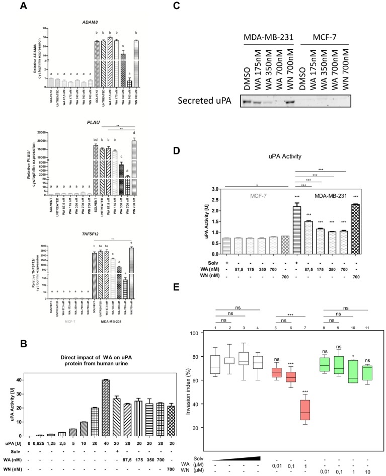
Figure 5. Decreased invasiveness of WA-treated MDA-MB-231 cells is associated with changed expression of ECM remodeling and pro-inflammatory genes.
(A) Effect of WA, WN on ADAM8, PLAU and TNFSF12 gene expression in MCF-7 and MDA-MB-231 cell lines normalized to cyclophilin housekeeping gene and relative to DMSO-treated MCF-7 sample (2-ΔΔCt) as determined by real-time quantitative PCR. Bar graphs represent relative mRNA (mean ± SEM) levels of three independent experiments. (B) Direct effect of WA, WN or DMSO on activity of uPA protein from human urine. Bar graphs represent mean ± SEM uPA activity of two independent experiments (C) Effects of DMSO, WN or WA at three different concentrations on uPA protein levels present in cell-conditioned medium was evaluated by Western blot. A representative blot picture of two independent experiments is shown. (D) The enzymatic activity of uPA in the cell-conditioned medium of MCF-7 and MDA-MB-231 cells treated with solvent, WA or WN as determined by CHEMICON colorimetric assay. Bar graphs represent mean ± SEM uPA activity of three independent experiments. (E) 24-hour collagen type-I invasion assay of MDA-MB-231 cells treated with DMSO, WA or WN. A box plot representing scored normalized invasion indexes of two independent experiments is shown. Within the frame, statistical significance is indicated for comparisons of treatment to the corresponding solvent condition. Above the frame, statistical significance is indicated for comparisons within a compound group.

