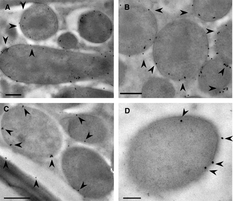
Figure 3.
Ultrastructural localization of AsE246 and DGDG on the symbiosome membrane. A and B, Immunogold detection of anti-AsE246 (immunogold signal appears as black dots; indicated by black arrowheads) in wild-type nodules. The 10-nm gold particles are present over the symbiosome membranes. C and D, Immunogold detection of anti-DGDG (immunogold signal appears as black dots; indicated by black arrowheads) in wild-type nodules. The 10-nm gold particles are present over the symbiosome membranes. Bars = 200 nm (A, B, and D) and 500 nm (C).

