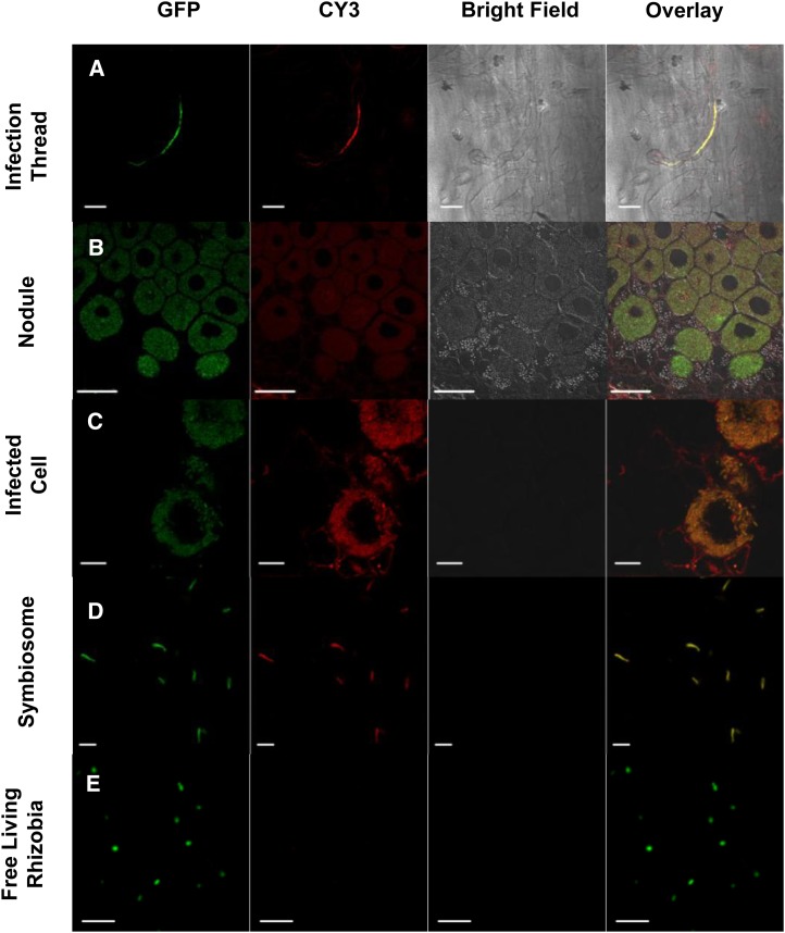
Figure 4.
DGDG is localized on the plasma membrane and colocalized with the symbiosome in infected cells. Immunolocalization of DGDG over the infected roots of Chinese milk vetch induced by M. huakuii 7653R shows colocalization with GFP-labeled 7653R in ITs and nodules but not with free-living M. huakuii. A, Immunofluorescence of DGDG on the IT; the rhizobia are expressing GFP, and the signal for DGDG is revealed as red dots (secondary antibody tagged with CY3). CY3 signal is detected both on root hair plasma membrane and colocalized with rhizobia (yellow color; in the overlay). B and C, Immunolocalization of DGDG on nodules. The signal shows in both infected and uninfected cells and colocalizes with rhizobia in the infected cells. D, Immunolocalization of DGDG in crushed nodules, which were fixed in 4% (w/v) of freshly depolymerized paraformaldehyde in 1× PBS. The signal shows that DGDG is localized on the symbiosome. E, Immunolocalization of DGDG on free-living M. huakuii rhizobia as a negative control shows that the free-living rhizobia do not contain DGDG. Bars = 20 μm (A and C), 50 μm (B), and 5 μm (D and E). [See online article for color version of this figure.]

