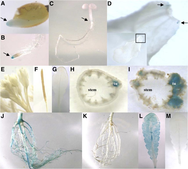
Figure 6.
Expression pattern of the SAD6 gene in Arabidopsis. A to D, Histochemical GUS assay indicated by the blue color in a 7-d-old seedling. Arrows point to GUS-derived blue color in the root apex of a germinating seedling (A), the root tip (B), the developing zone of the hypocotyl (C), and stipules at the base of the leaf petiole shown in closeup (D). E to I, GUS expression in a flowering plant: flowers (E), silique (F), and leaf (G) show no GUS activity; GUS-derived blue staining is seen in cross sections of an inflorescence stem with a young tumor (tu) 6 d post inoculation (H) and a mature tumor 21 d post inoculation (I) with A. tumefaciens strain C58. J to M, GUS expression in adult nonflowering plants treated with low oxygen (0.5% oxygen) or ambient oxygen concentrations for 24 h: strong GUS activity in a root (J) and a leaf (L) of a plant exposed to hypoxia and no GUS activity in a root (K) and a leaf (M) from a plant grown in normoxia. Representative images of three independent transgenic GUS-expressing lines of the T3 generation are presented.

