Figure 1.
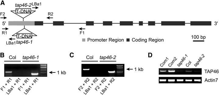
Molecular analysis of tap46 mutants. A, The TAP46 genomic structure and T-DNA insertion sites within the promoter sequence of the TAP46 gene. Dark boxes are exons, and the lines are introns. Primers that were used for PCR confirmation of the tap46-1 and tap46-2 mutants are marked by arrows. The primers tap46-F1 and tap46-R1 (F1 and R1) and tap46-F2 and tap46-R2 (F2 and R2) are specific for TAP46 DNA, and LBa1 (left border primer) is specific for the T-DNA sequence. B and C, PCR experiments demonstrate that the tap46-1 and tap46-2 mutants are homozygous. The primer combination for PCR reaction is shown below each lane. D, RT-PCR experiments demonstrate that the TAP46 transcript is reduced in the tap46-1 and tap46-2 mutants and elevated in complemented lines of the tap46-1 mutant. Com1 and Com2 indicate two complemented lines of tap46-1, respectively. Actin7 was used as the internal control.

