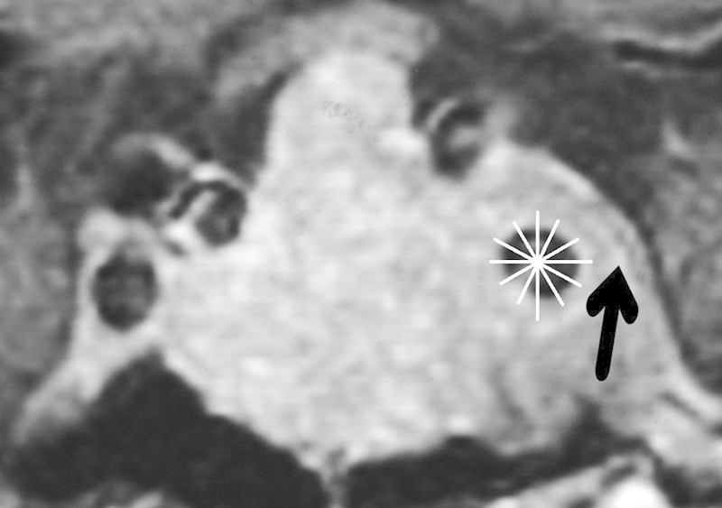
Fig. 3.

Gadolinium-enhanced coronal image demonstrating the division of the perimeter of the intracavernous internal carotid artery into twelfths like a clock face, used to score the degree of encasement. There is also bulging of the lateral wall of the cavernous sinus (arrow).
