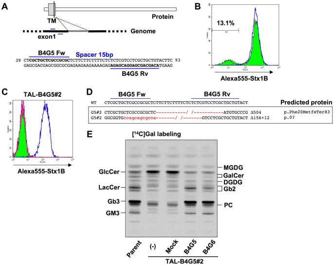
Figure 6. Isolation of a B4GalT5-deficient clone.
A, Target sites of TALEN-B4G5 pair (TAL-ModA-B4GalT5) in human B4GalT5 gene. The sequences are located in exon 1, which codes part of the transmembrane domain. The target sites are shown in bold. The numbers on the right and left of the sequence indicate the sequence numbers from the A of the translation initiation codon, based on B4GalT5 mRNA (accession number AB004550). B, Surface expression of StxRs on TALEN-B4GalT5–treated HeLa cells. HeLa cells were treated with TALEN-B4GalT5 (colored histogram with black line) or empty vectors (blue line), and the cells were stained with Alexa-555-Stx1B. C, Surface expression of StxRs on a TAL-B4GalT5 clone (TAL-B4G5#2). TAL-B4G5#2 cells were stained with Alexa-555-Stx1 B (colored histogram with black line) or not (magenta line) and HeLa-mCAT#8 cells were stained with Alexa-555-Stx1 B (blue line). D, Indel analysis of B4GalT5 gene in TAL-B4G5 clone (TAL-B4G5#2). The deletion is shown in red and its length specified on the right of the sequence. The predicted proteins are indicated based on the recommended description (see Materials and Methods) [50]. E, Metabolic labeling of lipids with radioactive galactose. TAL-B4G5#2 clone and B4GalT5- or B4GalT6-restored TAL-B4G5#2 cells obtained by retroviral vector–mediated overexpression were labeled with [14C]galactose for 16 h, and lipids extracted from the cells were separated by HPTLC. Radioactive image of an analyzed TLC plate is shown.

