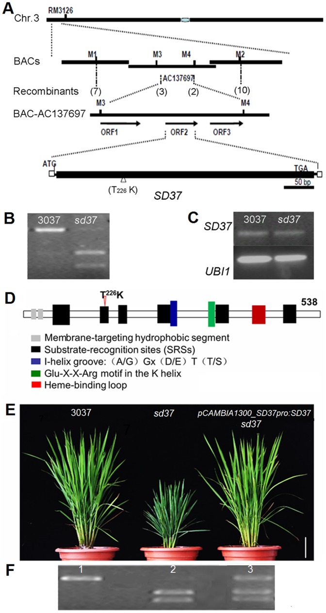
Figure 3. Map-based cloning of SD37.
(A) Physical mapping of SD37. The numbers in parentheses indicate the number of recombinants. SD37 was localized to BAC AC13769. The presumed ORFs were predicted using Gramene. White boxes indicate UTRs, and the black box represents the solitary exon. (B) Different sizes of the CAPS markers for 3037 and sd37 are shown using genomic DNA. PCR products of the SD37 CDS were amplified using the OE-F and OE-R primers (Table S3) and digested using AlwI. (C) SD37 expression in leaves from 3037 and the sd37 mutant were assessed using RT-PCR. Rice UBQ1 was used as an internal control. (D) Protein structure of SD37. The arrowhead indicates the point mutation in the SRS2 region. (E) Rescue of the sd37 phenotype with the pCAMBIA1300_SD37pro:SD37 construct. One representative complementation line (pCAMBIA1300::SD37) is shown. Bar = 10 cm. (F) CAPS marker detection in 3037 (lane 1), sd37 (lane 2), and a complementation line (lane 3). Samples were analyzed by agarose gel electrophoresis.

