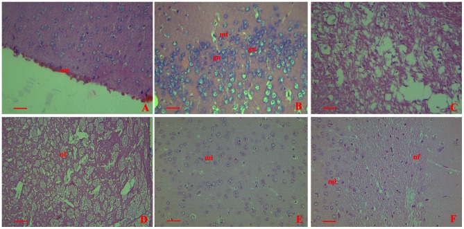
Figure 2. Histopathological changes shown by H&E staining in collected brains from mice infected with GD201008-001 at 20 h.p.i.
me = meninges;mi = microglia;gn = glial nodules;hc = Hippocampus;nf = nerve fibers. (A) Prominent meningeal hemorrhage and erythrocyte aggregation in the meninges (400×). (B) Microglial cells showing an increase in the number and volume. Glial nodules were distributed (400×). (C) Lytic and necrotic hippocampus (400×). (D) Nerve fibers showing severe damage (400×). (E) and (F) No histopathological changes in sham infection control (injected with PBS) (400×). Scale bar = 20 µm.

