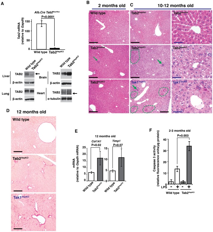
Figure 3. Hepatocyte-specific deletion of Tab2 induces late-onset fibrosis and sensitizes the liver to LPS-induced injury.
(A) Tab2 expression in the liver, lung, brain, and heart was determined by real-time PCR (liver) and immunoblotting (liver, lung, brain and heart) in hepatocyte-specific Tab2-deficient, Alb-Cre Tab2flox/flox (Tab2HepKO), and littermate or age matched control, Tab2flox/flox (wild type), mice. Wild type mice, n = 10; Tab2HepKO mice, n = 8. Means ± SEM and P values are shown. β-actin or α-tubulin is shown as a loading control. Arrows indicate the position of TAB2. (B) Hematoxylin and eosin staining of 2-month-old controls, including wild type and Tab2 heterozygous deletion, Alb-Cre Tab2flox/+ (Tab2HepHet), and Tab2HepKO liver sections. Scale bars, 200 µm. (C) Liver sections of 10- to 12-month-old control Tab2HepHet, Tab2HepKO and Tak1-deficient (Tak1HepKO) mice. Left panels show overall structure of liver. Arrows indicate immune cell infiltration, and dashed line areas contain highly eosinophilic cells. Scale bars, 200 µm (left panels), 40 µm (right panels). (D) Sirius Red staining of 12-month-old control, Tab2HepKO, and Tak1HepKO liver sections. Scale bars, 200 µm. (E) Fibrosis marker mRNA levels were determined by real-time PCR. Wild type mice, n = 6; Tab2HepKO mice, n = 3. Means ± SEM and P values are shown. (F) 2- to 3-month-old Tab2-deficient and control mice were treated with 10 mg/kg mouse weight LPS for 24 h. Protein extracts from the liver were subjected to caspase 3 assay. Wild type mice, n = 5; Tab2HepKO mice, n = 7. Means ± SEM and P values are shown.

