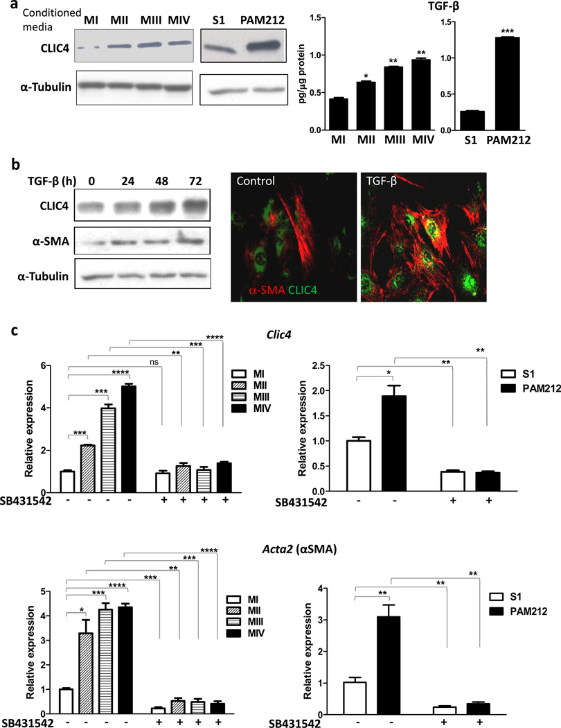
Figure 1.
Tumor cells induce CLIC4 and α-SMA expression in fibroblasts via TGF-β signaling. (a) Left Primary mouse dermal fibroblasts were treated with serum free conditioned media from either of the human breast cell lines MI, MII, MIII or MIV or the murine squamous cell lines S1 or PAM 212 cells. Expression of CLIC4 was analyzed by immunoblotting. Right TGF-β concentrations in conditioned media from human and mouse cell lines were determined by ELISA and normalized to total protein content. Data sets were compared for statistical significance with MI or S1(non-tumorigenic lines). (b) Primary dermal fibroblasts were treated for different time periods with TGF-β (10ng/ml) and immunoblotted (left) for CLIC4 and α-SMA. (Right) Co-immunofluorescence for CLIC4 (green) and α-SMA (red) in primary untreated fibroblasts or fibroblasts treated with TGF-β for 48h. (c) Primary dermal fibroblasts were treated with conditioned media as in A with or without pretreatment with the ALK5 inhibitor SB431542 (5µM). Expression of CLIC4 and α-SMA (gene name Acta2) was analyzed by real time PCR normalized to respective GAPDH levels and plotted as relative to MI or S1. Data sets were compared as indicated by lines for statistical significance.

