
Fig. 2.
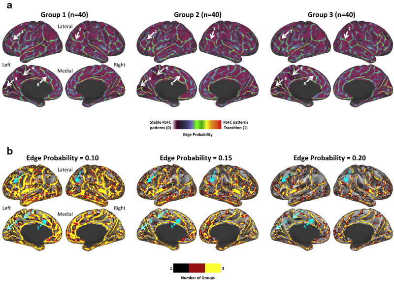
RSFC-Boundary Mapping parcellation reliably identifies locations of putative area borders. (a) RSFC-Boundary Mapping parcellations are highly similar across 3 independent groups of healthy young adults. A subset of locations is pointed out with arrows to highlight the high degree of similarity in parcellations. These locations include regions along the inferior and middle frontal gyri of the left hemisphere (1), a strong border separating angular gyrus from the middle-occipital gyrus in the right hemisphere (2), a strong border parallel to the calcarine sulcus in the medial occipital lobe (3), a strong border separating posterior extent of the cingulate gyrus from locations in the paracentral lobe (4), and a border which separates locations in the anterior cingulate gyrus from more dorsal regions of the medial frontal cortex (5). (b) The strongest RSFC-Boundary Mapping borders are consistent across groups. Independent conjunction images created by first thresholding each of the three group’s RSFC-Boundary Mapping parcellation maps from (a), binarizing the image, and summing the three images to demonstrate the consistency in parcellation features across groups. Three edge probability thresholds are depicted.