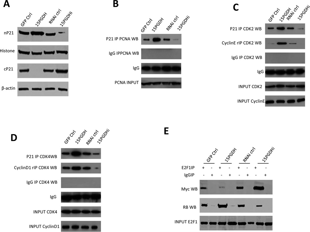
Figure 6. The effect of 15-PGDH signaling on p21 downstream molecules in HCC cells.
A. Western blotting for p21 in the nuclear fraction (nP21) and p21 in the cytoplasmic fraction (cP21) from Huh7 stable cell lines with altered 15-PGDH expression. Histone and β-actin were used as the internal control, respectively.
B. Co-immunoprecipitation and western blotting analysis using indicated antibodies in Huh7 stable cell lines with altered 15-PGDH expression. IgG IP was used as the negative control. PCNA western blotting was used as input control.
C. Co-immunoprecipitation and western blotting analysis using indicated antibodies in Huh7 stable cell lines with altered 15-PGDH expression. IgG IP was used as the negative control. rIP denotes repeat co-immunoprecipitation.
D. Co-immunoprecipitation and western blotting analysis using indicated antibodies in Huh7 stable cell lines with altered 15-PGDH expression. IgG IP was used as the negative control. rIP denotes repeat co-immunoprecipitation.
E. Co-immunoprecipitation and western blotting analysis using indicated antibodies in Huh7 stable cell lines with altered 15-PGDH expression. IgG IP was used as the negative control.

