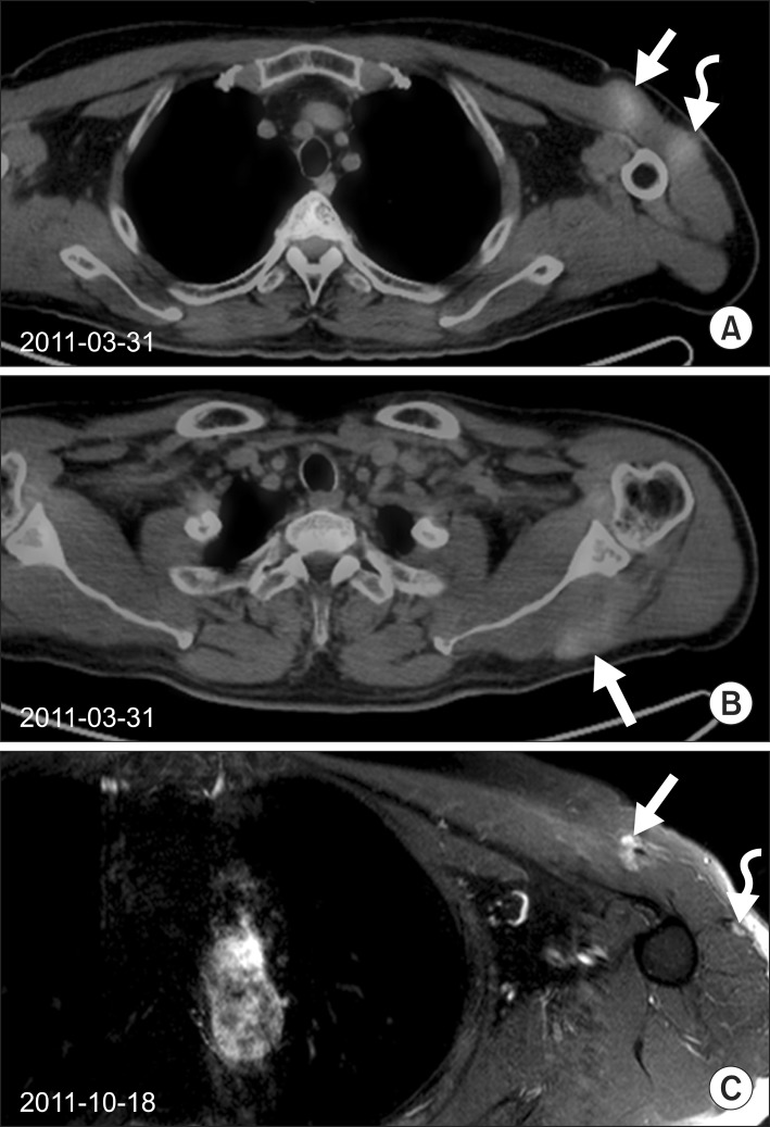
Fig. 1.
(A) Two well-enhanced intramuscular masses (3.5 cm × 1.8 cm; 2.2 cm × 1.9 cm) in the left pectoralis major (arrow) and left deltoid muscles (curved arrow) were visualized on PET-CT (maxSUV 2.57, 2.30) in March 2011. (B) An intramuscular mass along the left teres minor muscle was also visualized on PET/CT (maxSUV 2.00) in March 2011. Radiotherapy of 35 Gy in 14 fractions was delivered. (C) After radiotherapy of 35 Gy in 14 fractions, these masses were decreased in size (1.5 cm × 1.2 cm) markedly. PET/CT, positron emission tomography/computed tomography.

