
Fig. 1.
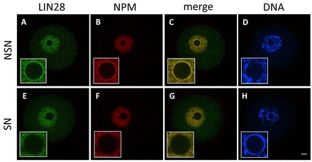
Cellular localization of LIN28 protein in fully grown mouse CD1 oocytes. Confocal immunofluorescence analysis of endogenous LIN28 in whole-mount oocytes. B23 (NPM) was used as a nucleolar marker. DNA was counterstained with DAPI. In NSN oocytes (A-D) LIN28 and NPM localize at the periphery of the nucleolus. During transition from NSN to SN configuration, both LIN28 and NPM gradually disappear from surface of the nucleolus and they are not detected there in SN oocytes (E-H). Insets show higher magnification of nucleus. Scale bar: 20 μm.