
Fig. 2.
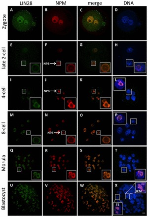
Cellular localization of LIN28 protein during mouse preimplantation development. Confocal immunofluorescence analysis of endogenous LIN28 in whole-mount embryos. B23 (NPM) was used as a nucleolar marker. DNA was counterstained with DAPI. An enlarged view of the boxed regions show that LIN28 (green) colocalizes with NPM (red). (A-D) In zygotes, homogenous nuclear signals of both LIN28 and NPM were observed. No enrichment at the periphery of nucleolar precursor bodies (NPBs) was observed for either marker. (E-P) At the early and mid 2-cell stage, LIN28 localizes at the periphery of nucleolar precursor bodies (NPBs) (arrow), where it colocalizes with NPM (E-H). LIN28 remains colocalized with NPM at NPBs (arrow) at the 4-cell (I-L) and 8-cell (M-P) stages. (Q-T) As NPBs have transformed to mature nucleoli in morula embryos, LIN28 remains nucleolar, as confirmed by the colocalization with NPM. (U-X) At the blastocyst stage, the localization of LIN28 in the nucleolus is concordant with NPM. LIN28 was present in both TE and ICM cells. Scale bar: 20 μm.