
Fig. 3.
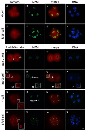
Cellular localization of a LIN28-Tomato fusion protein in early mouse embryos. Confocal immunofluorescence analysis of LIN28-Tomato fusion protein in whole-mount embryos following microinjection of mRNA, in vitro culture and fixation. B23 (NPM) was used as a nucleolar marker. DNA was counterstained with DAPI. As control, zygotes were microinjected with mRNA coding only for tdTomato protein. (A-H) Tomato expression is throughout the cytoplasm and nuclei in 4-cell embryos (A-D) and in 8/16 embryos (E-H). (I-L) At the 2-cell stage, the LIN28-Tomato fusion protein signal appears at distinct cytoplasmic foci that are formed around the nucleus (arrows). NPM staining is restricted to the nucleoplasm. (M-P) Focal plane of one blastomere of a late 2-cell embryo, where LIN28-Tomato localizes to one NPB, which is stained by NPM at the periphery (arrows). (M′-P′) Focal plane of second blastomere of the same late 2-cell embryo, where LIN28-Tomato signal is absent at presumptive NPBs, which lack NPM staining at periphery. (Q-T) At the 4-cell stage, LIN28-Tomato colocalizes with NPM at NPBs (an enlarged view of the LIN28-Tomato signal is shown in the boxed region). (U-X) In 8/16 embryos, LIN28-Tomato associates with mature, differentiated nucleoli. Scale bars: 20 μm in A-X; 10 μm in inset in U.