
Fig. 7.
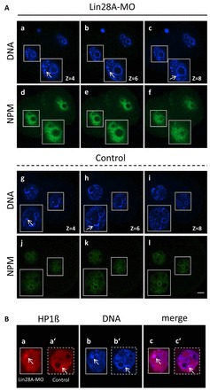
NPM expression and chromocenter formation following LIN28 depletion. Confocal immunofluorescence analysis of NPM (green) and HP1 (red) in whole-mount embryos. DNA was counterstained with DAPI. (Ad-f) Different optical slices illustrate that NPM is distributed throughout the nucleoplasm and is not enriched at the periphery of presumptive NPBs in 2-cell arrested embryos. (Aj-l) In control late 2-cell embryos, NPM is enriched at the periphery of NPBs. (Aa-c,g-i) Heterochromatin and chromocenter clustering is brightly stained by DAPI (arrow). Heterochromatic foci are outside (Aa,b,g) or in association (Ac,h) with NPBs (arrow). (Ba-c′) Chromocenters were strongly enriched in HP1. An enlarged view of a single optical slice shows that HP1 (red) colocalizes with condensed DNA (arrow) in 2-cell arrested (Ba-c) and control (Ba′-c′) embryos. Scale bar: 20 μm.